


Collective movement of cells viewed through the lens of fluid mechanics
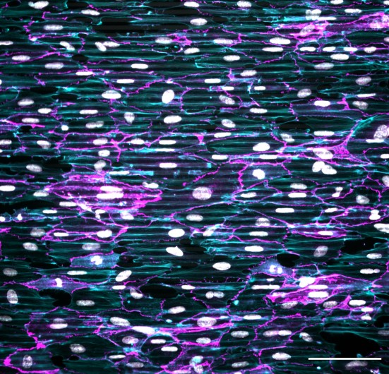 Cells are placed on a microgrooved substrate. Nuclei are coloured in white. Internal actin skeletons that follow the orientation of the grooves are coloured in cyan. Junctions between cells are coloured in magenta (scale 100 micrometres).
Beginning with embryonic development and throughout our lives, the cells in our bodies are in incessant motion. Cells can move as individuals or in coordinated groups, a process called “collective migration”. A question that has long fascinated scientists is how cells move and how they are guided to reach their target location. Historically, such questions have been principally viewed from a biochemical perspective, with the identification of chemical factors involved in cell migration.
Over the past couple of decades, however, the importance of physical factors in collective cell migration has been recognized and has attracted growing interest from physicists who have demonstrated similarities with fluid mechanics. Indeed, groups of cells moving collectively can exhibit swirling-like motions akin to turbulent fluid flow. In addition, scientists have observed that 2D confinement of cell assemblies using selectively adhesive areas of specific shapes can generate original patterns of cellular movement such as rotation or bidirectional motion.
In a study published in Nature Communications, a team of researchers from Hydrodynamics Laboratory at Ecole Polytechnique (LadHyX*) focused on a more physiological setting. Rather than imposing global confinement of the cells, they cultured them on substrates composed of parallel arrays of microgrooves that impose constraints on each cell’s surface and lead to cellular elongation and alignment in the direction of the grooves. This type of confinement is more representative of the in vivo situation, where cells are guided and oriented by the fibers of the extracellular matrix to which they adhere. By recording the collective movement of vascular endothelial cells (the cells lining the inner surfaces of our blood vessels) on these microgrooves, a specific pattern of movement was observed. It is characterized by periodic antiparallel streams, i.e. corridors of cells that alternatively move from right to left and left to right :
“To explain the emergence of this surprising pattern, we teamed up with a physicist who developed a mathematical model where the cells are considered as an active fluid” says Claire Leclech, first author of the study. This model is able to predict the formation of the antiparallel cell streams observed experimentally. Finally, the researchers demonstrated that other external constraints that orient the cells also lead to this collective movement, for example by subjecting the cells to a unidirectional fluid flow. By identifying the minimal ingredients necessary to obtain this particular pattern, this study highlights once again the interest of bringing biology and physics together.
*LadHyX: a joint research unit CNRS, École Polytechnique - Institut Polytechnique de Paris