 Support l'X
 See the structure of collagen in 3D
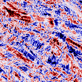
 (a): principle of circular dichroism measurements in SHG on collagen fibrils oriented out of the imaging plane and of different polarities (pointing upwards in green and downwards in violet); (b): image of circular dichroism in SHG of a cross-section of human cornea.
Studying the structure of collagen
Collagen fibrils are an essential component of biological tissues and their three-dimensional distribution is decisive for obtaining specific tissue functions. Thus, the dermis of the skin is opaque and resilient because it is composed of a disorderly tangle of large fibrils, while the cornea is transparent and rigid because it is made up of fine, well-ordered fibrils in a plywood-like structure.
It is therefore essential to precisely characterize the three-dimensional distribution of these collagen fibrils in order to better understand the link between the structure of a tissue and its biological function, but also to be able to reproduce this structure in vitro and obtain high-performance biomimetic tissues for transplants or biomedical studies.
In the case of pathologies, this characterization is also essential: the tissues are usually remodeled and the collagen exhibits a more disordered structure (for example skin scars). Being able to finely characterize these structures should allow to improve the diagnosis or knowledge of these pathologies.
SHG: a proven technique
The reference technique for imaging collagen in 3D is Second Harmonic Generation microscopy or "SHG", which is part of the optical processes called "multiphoton". These SHG signals make it possible to visualize very precisely the collagen fibrils in intact biological tissues.
By playing on the polarization state of the light, it is also possible to highlight the direction of the fibrils in the imaging plane. However, fibrils oriented out of the imaging plane emit a signal that is very weak and poorly detected.
Circular polarizations to go further
For the first-time, a research team led by Marie-Claire Schanne-Klein, CNRS research director at the Optics and Biosciences Laboratory*, has succeeded in efficiently visualizing collagen fibrils located out of the imaging plane. To do this, they carried out measurements of circular dichroism in SHG, i.e. measurements of the normalized difference between SHG signals excited by right and left circular polarizations. Their work was published in the Optica journal of the Optical Society OSA this month.
This method is made possible by the chirality of collagen molecules (because they are helical). For this reason, it requires to model SHG signals beyond the electric dipole approximation, i.e. to consider the magnetic contributions that were previously neglected.
The results of the numerical simulation, in agreement with the images of human corneas and collagen matrices studied in the laboratory, show that this method makes it possible to probe the degree of polarity of fibrils outside the imaging plane, i.e. to measure a new collagen disorder parameter in 3D.
Learn more: read the publication
*LOB, a UMR CNRS-Polytechnic School-Inserm