 Support l'X
 How ribosomes in archaea enter hibernation
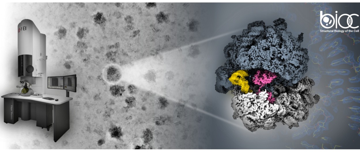 The scientists used microscopes at the Cimex facility at École Polytechnique to image the ribosomes and reconstruct them in 3D
Ribosomes play a key role: they assemble proteins from genetic information in all living organisms. These proteins then perform all the functions necessary for life. A ribosome consists of two subunits: a small one responsible for reading the messenger RNA that contains the genetic information, and a large one that assembles the corresponding protein. Thousands of these ribosomes are present in every cell; they are massive and consume a great deal of energy.
But when energy becomes scarce, a way must be found to shut down the ribosomes and protect them from degradation until conditions become favorable again. In an article published in Nature Communications, the translation mechanisms team at BIOC reveals a protein capable of putting archaeal ribosomes into “hibernation.”
Archaea are single-celled organisms distinct from bacteria (which are also single-celled and lack a nucleus) and closely related to eukaryotes (a group comprising all living organisms whose cells possess a nucleus). Recent studies show that they are found in all types of environments, even in the human microbiome! Like all living organisms, archaea are subject to environmental fluctuations and must adapt their energy consumption accordingly.
“The factors involved in ribosomal hibernation were already well understood in bacteria and eukaryotes. But before we began our work, none had been identified in archaea,” explains Clément Madru, assistant professor in the Department of Biology at École Polytechnique and co-first author of the article alongside Gabrielle Bourgeois (BIOC) and Rémi Dulermo (Ifremer).
HibA: A Key Protein for Putting Ribosomes into Standby Mode
The scientists subjected Pyrococcus abyssi archaea to temperature and oxygen stress. This hyperthermophilic archaeon normally grows in the absence of oxygen, in the abyssal trenches of the Pacific Ocean. They then purified the ribosomes from these stressed cells. Finally, using the cryo-electron microscope at the École Polytechnique’s Interdisciplinary Microscopy Center (CIMEX), a very large number of ribosomes were examined in minute detail, allowing their three-dimensional structure to be recontructed, which is one of BIOC’s specialties.
Most of the ribosomes in the sample were bound to a molecule whose function was previously unknown, named HibA. One part of HibA, similar to a molecule already known in other organisms, can bind to the small subunit of the ribosome in place of messenger RNA. The other part binds to functional sites on the large subunit.
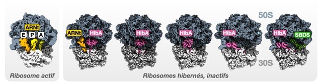
Caption: Ribosomal structures from the archæa Pyrococcus abyssi. On the left, an active ribosome associated with transfer RNAs (tRNAs) involved in protein synthesis. On the right, the hibernation factor HibA bound to ribosomes observed in various states. HibA binds to the interface between the large subunit (50S) and the small subunit (30S).
These bonds prevent the ribosome from functioning, effectively putting it into hibernation. “It also serves to hold the two subunits together, preventing the ribosome from dissociating. This helps protect it from degradation while it waits for better conditions,” says Clément Madru. “Furthermore, HibA can occupy different positions within the ribosome. This is the first time a hibernation factor has been identified with multiple binding sites.”
The data also show that HibA can bind to molecules that supply energy to the cell. “This may be linked to its ability to block ribosomes, but it is still just a hypothesis we are working on,” notes the researcher. Beyond deciphering cellular mechanisms, the comparative study of ribosomes present in different types of organisms sheds light on the overall evolution of life.
Reference :
Madru, C., Bourgeois, G., Dulermo, R. et al. A family of ribosome hibernation factors widespread in Archaea. Nat Commun (2026). https://doi.org/10.1038/s41467-026-72341-8
*BIOC: a joint research unit CNRS, École Polytechnique, Institut Polytechnique de Paris, 91120 Palaiseau, France
The collaboration includes scientists from BIOC, Ifremer, the University of Toulouse, Université Paris-Cité, and Institut Pasteur.