 Support l'X
 When light stiffens cells
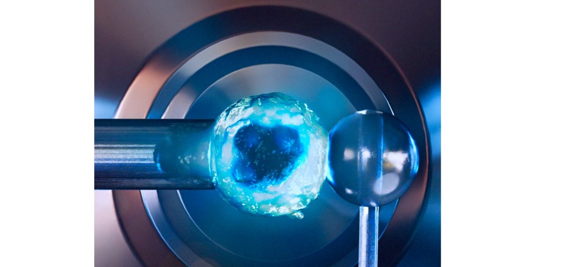 A lymphocyte held by a micropipette is brought close to a glass bead in order to measure its rigidity. Artist's impression of an experiment carried out at LadHyX. Image by Julien Husson (https://cellmechanics.jimdofree.com/).
During an experiment using a microscope to observe the behavior of immune cells, Julien Husson and his team noticed something unexpected: the cell, a lymphocyte, appeared to become inert, as if hardened. During these experiments, the scientists added fluorescent molecules capable of binding to the calcium. When illuminated with blue light, these molecules emit a signal proportional to the amount of calcium, thus allowing cellular activity to be measured.
Phototoxicity and stiffening
This fluorescent microscopy technique is widely used in biology, and phototoxicity was already known, but not the stiffening effect. "Scientists using fluorescence microscopy are aware of this phototoxicity, and observation protocols take it into account in order to minimize it. However, the link between this phototoxicity and a change in cellular mechanics had not been studied in detail," explains Julien Husson, a researcher at the Hydrodynamics Laboratory (LadHyX*), who sought to learn more through a collaboration**.
In the experiments he designs at LadHyX, mechanical properties can be measured at the cellular level using micropipettes. A lymphocyte (a type of immune cell) is gently sucked onto the tip of a micropipette and then pressed against a thin glass rod. The bending of the rod allows the force applied to be measured, and by measuring the displacement of the micropipette, scientists can deduce how much the cell has deformed. They can then quantify the rigidity of the cell. Under normal circumstances, a lymphocyte is very soft.
When the interior of a cell is loaded with fluorescent molecules, the rigidity of the cell does not change. However, when the cell is illuminated with light, things change radically, and sometimes the cell becomes up to ten times more rigid! The cell becomes hard and loses its ability to act. “We tested different fluorescent molecules and different types of cells, from white blood cells to endothelial cells: there is always a stiffening compared to experiments carried out on control cells,” adds Julien Husson.
Role of reactive oxygen species
What causes this stiffening? The authors suggest that it is probably due to chemical species created when light activates the fluorescent molecule. This illumination correlates with the production of reactive oxygen species, which have the ability to form strong (covalent) bonds with the molecules around them. “Our hypothesis is that these bonds are responsible for cell hardening. To take this further, we would need to demonstrate the local presence of covalent bonds during this process,” explains the researcher.
Julien Husson sees potential applications for this research. For example, it could benefit a cancer therapy called photodynamic therapy. This involves injecting a molecule into cancer cells which, when exposed to light, generates reactive species that cause the tumors to die. Measuring rigidity could help evaluate the effectiveness of this method and possibly adjust the doses.
Article's reference :
Eva Gonzalez, Jana El Husseiny, Finn Bastian Molzahn, Tiffany Campion, Hadrien Jalaber, Stéphanie Dogniaux, Pierre-Henri Puech, Oliver Nüsse, Laure Gibot, Julien Husson, Stiffening cells with light, Cell Reports Physical Science, 2025, https://doi.org/10.1016/j.xcrp.2025.10299
*LadHyX: a joint research unit CNRS, École Polytechnique, Institut Polytechnique de Paris, 91120 Palaiseau, France
**Laboratoire Adhésion et Inflammation, Aix Marseille Université UM61, CNRS UMR 7333, Inserm U1067, Marseille ; Laboratoire Softmat, Université de Toulouse, CNRS UMR 5623, Toulouse ; Institut de Chimie Physique, CNRS UMR 8000, Université Paris-Saclay, Orsay ; Analyse integrative de l'activation des lymphocytes T, Institut Curie-PSL Research University, INSERM U932, Paris